Histiocytomas In Dogs: Diagnosis And Treatment Of These Benign Tumors
Histiocytomas in Dogs
If your young dog suddenly develops a red, ulcerated lump, they might have a histiocytoma. We spoke with Dr. Shagufta Mulla, a veterinarian with a DVM degree from Colorado State University with 20 years of experience as a small animal veterinarian, who told us that while the overwhelming majority are benign, a small percentage of histiocytomas are malignant. Histiocytomas can resemble skin growths that are malignant, including melanomas and mast cell tumors. That's why it's important to take your dog to the veterinarian for an examination if you find a lump on your dog's skin.
 WilleeCole/iStock/Getty Images
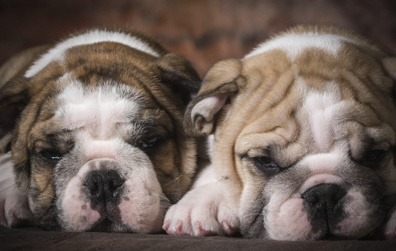
Canine cutaneous histiocytomas
Canine cutaneous histiocytomas
Histiocytomas are common and pet owners will typically notice a single, hairless lump that is raised and often has ulceration. They are typically less than 2.5 centimeters in diameter. Because of their appearance, they've been dubbed "the button tumors." They originate from a type of lymphocyte (or white blood cell) called Langerhans cells, which are part of your dog's immune system. These cells are commonly found in the skin.
Histiocytomas most often show up on the front half of the dog's body, especially the face, neck, ears and legs, but not always. When you palpate (feel with your hands) a lump and it slides around easily, it's probably benign as compared with malignant tumors, which usually don't move when palpated. "A notable exception is the mast cell tumor, which is a common cancer in dogs that can sometimes resemble a benign lipoma," says Dr. Mulla. Your dog might scratch at a histiocytoma, causing bleeding and secondary infection. Occasionally, the lymph node that's closest to this inflammatory process might swell.
 Jennifer_Sharp/iStock/GettyImages
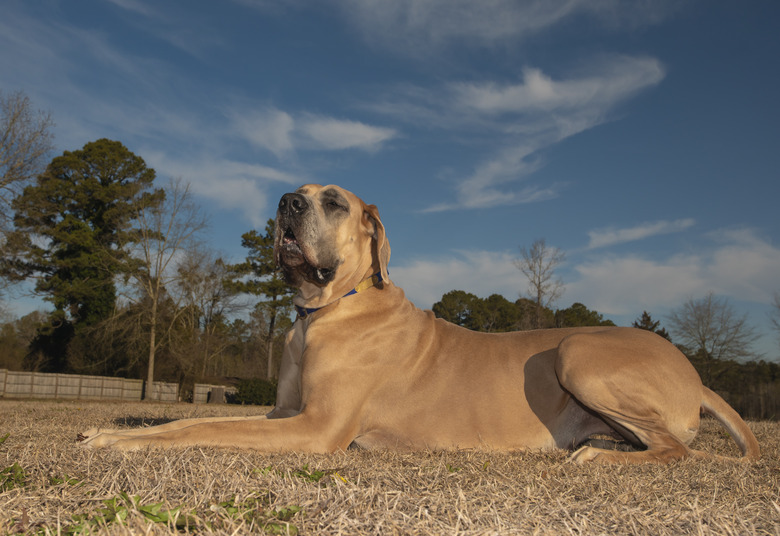
Dog breeds more prone to histiocytomas
Dog breeds more prone to histiocytomas
The cause of histiocytomas is unknown but these benign tumors appear more often in certain breeds. These include cocker spaniels, great Danes, dachshunds, boxers, Shetland sheepdogs, bull terriers, Labrador retrievers, greyhounds, American Staffordshire bull terriers, flat-coated retrievers, bulldogs, Scottish terriers, Boston terriers and Shar-Peis. Benign histiocytoma growths generally appear in young animals, usually 3 years old or younger. Malignant histiocytomas most often occur in Swiss mountain dogs, Rottweilers and golden retrievers, although any dog is susceptible.
Cutaneous histiocytoma diagnosis and treatment in dogs
Cutaneous histiocytoma diagnosis and treatment in dogs
To get a definitive diagnosis, your veterinarian will initially use fine needle aspiration (FNA) to take cells from the tumor. If needed, they will biopsy the lump or, in certain cases, perform surgical excision. With FNA, your veterinarian prepares slides for cytology, which they may look at under a microscope in-house and/or submit to a laboratory. "With fine needle aspirate, sometimes it's possible to diagnose histicytomas in-house under the microscope, without sending the slides to a pathologist," Dr. Mulla says. With biopsy or surgical removal of the lump, they'll send the sample to a veterinary pathologist for histopathology to obtain a diagnosis.
If the initial fine needle aspiration shows that the tumor is benign, no further treatment like surgery is necessary. However, if your dog is bothering the lump because it's itchy or bleeding, your veterinarian can remove it. "They're not usually excised unless they're causing a problem, for example, your pet won't leave it alone or the lump keeps bleeding," explains Dr. Mulla. They may also have you try an Elizabethan collar and a topical anesthetic or ointment to calm the itch and ulceration. Unlike many other forms of benign tumors, histiocytomas often disappear on their own, usually within two to three months after their appearance.
 gsagi/iStock/GettyImages
Canine malignant histiocytomas
Canine malignant histiocytomas
Although the overwhelming majority of histiocytomas are benign and regress on their own, some turn out to be cancerous. Malignant histiocytomas generally appear in middle-age canines. A skin tumor might be a visible sign of disease that has already spread inside the body, especially to the lymph nodes, spleen, bones, kidney and liver.
Symptoms of cancerous histiocytoma include weight and appetite loss, lethargy, breathing difficulties, fever and neurological problems. If your dog is diagnosed with malignant histiocytoma, treatment consists of combination chemotherapy, cell therapy and steroids. This regimen has kept affected dogs alive from several months to six years.
The bottom line
The bottom line
Histiocytomas are benign skin tumors that often appear on dogs 3 years of age and younger. They usually regress on their own within two or three months after appearing. Unless they're causing a problem for your dog, they won't be surgically removed. An Elizabethan collar and ointment can be tried to keep your pup more comfortable. Sometimes older dogs can get a cancerous form of histiocytoma called malignant histiocytoma.